
Key Takeaways
- The NIH BICAN consortium published a 26-paper atlas profiling 25.5 million cells across eight basal ganglia structures in humans, macaques and marmosets.
- The atlas identifies 61 cell types conserved across 76 million years of evolution and localizes disease risk to specific cell populations.
- The core human reference rests on six donated brains, and the atlas is a reference scaffold for disease research rather than a treatment.
The brain region that turns thoughts into actions
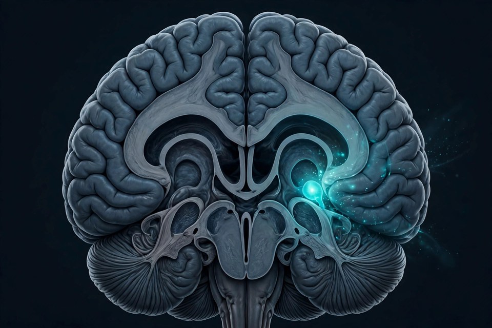
Deep in the brain sits a cluster of structures most people have never heard of and nobody can live without: the basal ganglia. They take the brain's intentions — move, choose, repeat, stop — and gate them into action, and they sit at the centre of Parkinson's disease, Huntington's disease, schizophrenia, bipolar disorder and addiction. This week, an international consortium published the most detailed map of the region ever made: 26 papers profiling 25.5 million cells from humans, macaques and marmosets.
The project, the U.S. National Institutes of Health's BICAN consortium, combined gene expression, epigenetics and spatial mapping across eight basal ganglia structures. Its headline product is a shared taxonomy: 61 cell types that appear in all three primate species — cell types conserved across 76 million years of evolution.
Why a parts list changes the science
A map like this earns its keep in the disease column. The atlas's cell-by-cell resolution lets researchers localize risk: genetic risk for psychiatric and neurodegenerative diseases does not spread evenly across the basal ganglia but concentrates in specific cell populations — including a previously under-characterized type of striatal neuron the team singles out. Developing-brain maps in the same package found that the neurons which mature in this region carry elevated genetic risk for schizophrenia and bipolar disorder.
Other papers in the set found structure where microscopes see none: the striatum, the region's largest structure, divides into six molecular zones with distinct gene signatures — zones whose signatures blur with age, especially in areas hit by Huntington's and Parkinson's. And a study of 178 donors showed that the cellular make-up itself, especially of glial cells, varies from person to person — a warning that no single "standard brain" exists.
The honest limits
Two caveats keep the achievement in proportion. The core human reference rests on just six donated brains — an extraordinary depth of data per brain, but a narrow slice of humanity. And an atlas is a scaffold, not a cure: as one consortium scientist put it, it is a framework "on which we can now fairly readily complete the rest of the brain." What it gives medicine is the dictionary — the named cell types, in their places, against which diseased brains can now be compared cell by cell.
The Twikup Take
Brain science has long described diseases by the region that fails. This atlas starts describing them by the cells that fail — 61 ancient cell types, mapped at last. Parkinson's and Huntington's research just got a much sharper address book.
Image: Illustrative image.